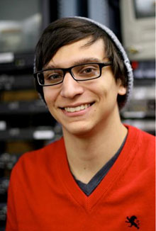
| As he tailors one of the world's finest imaging instruments to tackle one of science's most baffling challenges, Tom Flores feels like he's playing a microscopic game of Where's Waldo. | |||
| In the children's books by Martin Handford, readers pore over illustrations crammed with hundreds of people to search for Waldo and his trademark red-and-white-striped shirt. | |||
| Flores, a junior majoring in physics, is on a quest for something more elusive—the tiny carbon nanotube. | |||
Carbon nanotubes measure 1 to 5 nanometers in diameter. One nanometer is a billionth of a meter, or between one ten-thousandth and one hundred-thousandth the thickness of a human hair. With unmatched strength, stiffness and hardness, and length-to-diameter ratios of as much as millions to one, CNTs have potential in medicine, energy and many other applications. But their infinitesimal size makes it difficult to find and observe CNTs. While Waldo hides behind people, CNTs conceal themselves among bumps, nicks, specks of dust and other imperfections on a microscope slide. They reveal their presence by emitting infrared light when a light source is directed at them. An ultrathin plane of focus Flores began studying CNTs last spring with Slava Rotkin, associate professor of physics, and continued last summer in the physics department's Research Experience for Undergraduates program. Funded by the National Science Foundation, the program enables students to do a 10-week paid internship alongside a faculty member. Lehigh's REU program, with more than two decades of NSF funding, is one of the nation's oldest. In the past five years, an average of 25 to 28 students, roughly one-third from Lehigh, have taken part in the internship. Flores and two graduate students—Massooma Pirbhai and Tetyana Ignatova—study CNTs with a custom-made NTEGRA-Spectra recently acquired by Rotkin and Richard Vinci, professor of materials science and engineering. The instrument pairs an optical microscope with an atomic-force microscope (AFM), whose needle-like probe scans a surface and records its topographical features. | |||
| Flores and his colleagues combine AFM with an optical imaging technique called total internal reflection fluorescence. | |||
| "TIRF is a form of photoluminescence," says Flores. "You excite an object so that it gives off light, which provides information about the object and its properties. | |||
| "TIRF can excite an object in an extremely thin plane. We study single-walled CNTs, which are 1 nm in diameter. Our plane of focus has to be very thin; if not, we get luminescence from impurities near our sample." | |||
| A unique integration of microscopy techniques | |||
| Flores uses the AFM probe tip to locate the position of CNTs on a sample. | |||
| "We produce an AFM topographical image that shows us where we need to focus. The resolution of that image is limited only by the diameter of the tip. This is much better than you can do with an optics probe. | |||
| "Our project is like a game of Where's Waldo. We're trying to find a tiny object in a giant sample. We have to combine information from the AFM about physical characteristics—shape and size—with information supplied by TIRF about how light interacts with the sample." | |||
| Only one other research group in the U.S., says Flores, integrates AFM and TIRF in a setup exactly the same as Lehigh's. Combining the two techniques requires resourcefulness. To achieve optimum focus and illumination, Flores and his colleagues have had to modify the sample stage and lenses of the optical microscope. | |||
| "Our overall goal is to find and examine CNTs and characterize their properties so that engineers can find applications for them. | |||
| "We don't have images of CNTs yet, but we have produced images of polyethylene beads with dyes that emit light at various wavelengths. | |||
| "So we know our system is working." | |||
| Source: By Kurt Pfitzer, Lehigh University |

